


Semen
Semen is a suspension containing spermatozoa cells and various components secreted by accessory glands located in the male reproductive organ. Generally, the components of semen can be categorized into two parts: spermatozoa cells and the fluid phase in semen, often referred to as seminal plasma. Spermatozoa are produced by the testes, while the seminal plasma is produced by the vesicular and prostatic glands (Garner and Hafez, 2000).
In cattle, 90% of the semen components are dominated by seminal plasma, which serves as a medium for carrying sperm from the male reproductive tract to the female reproductive tract. This function is facilitated because semen contains many buffers and sources of nutrients for spermatozoa, both directly usable ones like fructose and sorbitol, as well as indirectly through Glycerylphosphorylcholine (GPC) (Toelihere, 1993).
Spermatogenesis
Spermatogenesis is a process involving the multiplication and differentiation of germ cells to produce male gamete cells (spermatozoa). This process occurs in the testes, specifically in the seminiferous tubules. Spermatozoa are released from the apical pole of Sertoli cells located in the seminiferous tubules (Illustration 1). Generally, spermatogenesis is divided into three stages: 1) Spermatocytogenesis, 2) Meiosis, and 3) Spermiogenesis. Spermatocytogenesis begins with the proliferation of spermatogonia cells. In this phase, spermatogonia produce two types of diploid cells: cells for renewing the stock of spermatogonia and proliferating spermatogonia that transform into primary spermatocytes (Garner and Hafez, 2000; Auger, 2018).
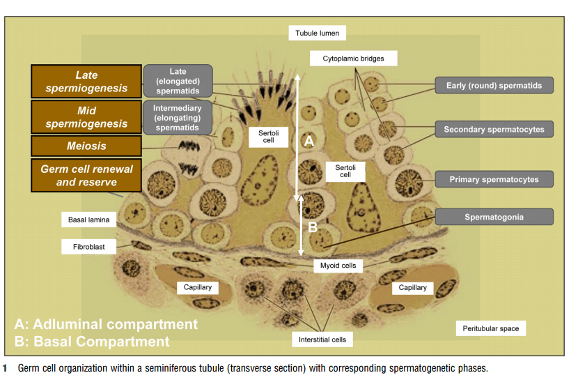
Illustration 1. Seminiferous tubules as the site of spermatogenesis (Auger, 2018).
The meiotic phase begins when primary spermatocytes pass from the blood-testis barrier to the lumen of the seminiferous tubules. Meiosis is divided into two stages: meiosis I, involving reduction of chromosome number and separation of homologous chromosomes, followed by meiosis II, involving separation of sister chromatids that eventually become individual chromosomes. Meiosis II results in spermatids. The spermatocytogenesis process up to the formation of spermatids takes about 40 days. The final stage is spermiogenesis, in which spermatids differentiate into more complex forms called spermatozoa (Garner and Hafez, 2000; Staub and Johnson, 2018; Auger, 2018). The stages of spermiogenesis are illustrated in Illustration 2.
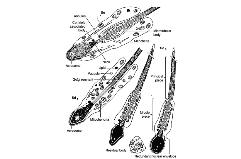
Illustration 2. Specific stages of spermiogenesis, from spermatid (Sa) transforming into spermatozoon (Sd 2) (Kretser, et al., 2018)
Male testes consist of 90% seminiferous tubules, while the remaining 10% comprises interstitial cells and connective tissue. After puberty, the testes develop rapidly, causing the seminiferous tubules to elongate and their diameters to increase. This increase in the volume of seminiferous tubules enhances the efficiency of spermatogenesis, resulting in a significant increase in the number of spermatozoa (Schenk, 2017). Inside the seminiferous tubules are spermatogonia cells up to spermatozoa cells. Additionally, Sertoli cells are present, which generally provide nutrition to spermatozoa but also serve as a blood-testes barrier, produce inhibin hormone, and convert testosterone hormone to estradiol 17β (estrogen). Interspersed among the seminiferous tubules are interstitial cells, including Leydig cells that produce testosterone, which is not only essential for spermatogenesis but also for the maturation of spermatozoa in the epididymis (in the form of dihydrotestosterone) and for increasing libido for mating (Susilawati, 2011). Spermatogenesis is also controlled by Follicle Stimulating Hormone (FSH) secreted by the anterior pituitary (Ismaya, 2014).
Under normal physiological conditions, beef cattle can produce about 4×10⁹ spermatozoa cells per day, while dairy cattle can produce about 7×10⁹ cells per day (Ismaya, 2014). However, this sperm production can be affected by external factors. Spermatogenesis is influenced by factors