


Semen
Semen is a suspension containing spermatozoa cells and various components secreted by accessory glands located in the male reproductive organs. Generally, the components of semen can be categorized into two: spermatozoa cells and the liquid phase within semen, often referred to as seminal plasma. Sperm is produced by the testes, while the seminal plasma is produced by the vesicular and prostatic glands (Garner and Hafez, 2000).
In cattle, 90% of the semen's composition is dominated by seminal plasma, which serves as a medium for carrying sperm from the male reproductive tract to the female reproductive tract. This function is efficiently carried out due to the presence of various buffers and nutrients for spermatozoa in the semen, including direct sources like fructose and sorbitol, as well as indirect sources like Glyceryphoricholine (GPC) (Toelihere, 1993).
Spermatogenesis
Spermatogenesis is the process of germ cell multiplication and differentiation aimed at producing male gametes (spermatozoa). This process occurs in the testes, specifically in the seminiferous tubules. Spermatozoa are released from the apical pole of Sertoli cells located in the seminiferous tubules (Illustration 1). Spermatogenesis is generally divided into three stages: 1) Spermatocytogenesis, 2) meiosis, and 3) Spermiogenesis. Spermatocytogenesis begins with the proliferation of spermatogonia. During this phase, spermatogonia produce two types of diploid cells: cells for replenishing the stock of spermatogonia and proliferating spermatogonia that transform into primary spermatocytes (Garner and Hafez, 2000; Auger, 2018).
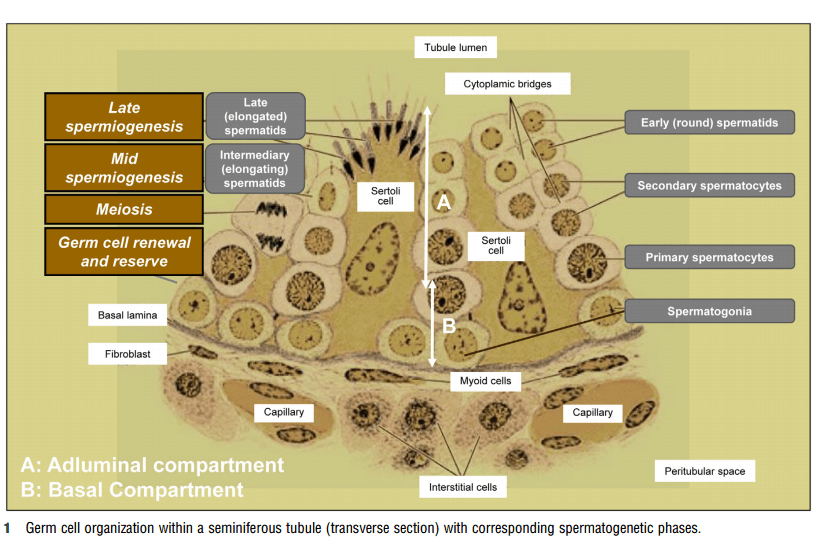
Illustration 1. Seminiferous tubules as the site of spermatogenesis (Auger, 2018).
The meiosis phase begins when primary spermatocytes traverse the blood-testis barrier toward the lumen of the seminiferous tubules. Meiosis consists of two stages: meiosis I, which reduces the chromosome number and separates homologous chromosomes, followed by meiosis II, which separates sister chromatids, ultimately resulting in individual chromosomes. Meiosis II produces spermatids. The process from spermatocytogenesis to spermatid formation takes about 40 days. The final stage is spermiogenesis, the process by which spermatids differentiate into more complex forms known as spermatozoa (Garner and Hafez, 2000; Staub and Johnson, 2018; Auger, 2018). The stages of spermiogenesis are depicted in Illustration 2.
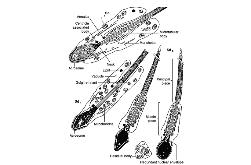
Illustration 2. Specific stages of spermiogenesis, from spermatid (Sa) to transformed spermatozoon (Sd 2) (Kretser, et al., 2018)
Male testes are composed of 90% seminiferous tubules, with the remaining 10% consisting of interstitial cells and connective tissue. After puberty, testes rapidly develop, resulting in elongation and thickening of seminiferous tubules. Increased tubule volume makes the spermatogenesis process more efficient, leading to a significant increase in the number of spermatozoa (Schenk, 2017). The seminiferous tubules contain spermatogonia cells up to spermatozoa, along with Sertoli cells, which provide nutritional support to spermatozoa and also act as blood-testis barriers. Leydig cells are located between seminiferous tubules and are responsible for producing testosterone, essential for spermatogenesis and the maturation of spermatozoa in the epididymis (in the form of dihydrotestosterone). Testosterone also increases libido for mating (Susilawati, 2011). Spermatogenesis is also controlled by Follicle Stimulating Hormone (FSH) secreted by the anterior pituitary (Ismaya, 2014).
Under normal physiological conditions, beef cattle can produce approximately 4 × 10⁹ sperm cells per day, while dairy cattle can produce around 7 × 10⁹ sperm cells per day (Ismaya, 2014). However, this sperm production can change due to external factors. Spermatogenesis is influenced by various factors, including environmental temperature and nutrition. Germinal epithelial cells are particularly sensitive to heat, especially during spermatid development. Heat radiation affects not only spermatid development but also spermatogonial division. Heat radiation before puberty has a significant impact on sperm cell production. Nutritional deficiencies can lead to testicular hypoplasia, affecting accessory glands and delaying puberty. Energy deficiency in feed can influence gonadotropin secretion, delaying sexual maturity, and resulting in reduced libido, semen volume, and quality (Susilawati, 2011).
2.3. Morphology of Spermatozoa
The function of spermatozoa is to deliver paternal genes (haploid) to the egg cell and initiate embryo development. To achieve this, the structure of spermatozoa has evolved to compartmentalized cells with specialized functions in each compartment. Generally, spermatozoa are divided into a head region and a tail (flagellum). The head can be further divided into an acrosomal region and a nucleus. The acrosomal region consists of secretory acrosomal vesicles and the plasma membrane that encloses the acrosome (Gerton and Vadnalis, 2018). This region contains acrosin, hyaluronidase, and other hydrolytic enzymes used in the fertilization process (Garner and Hafez, 2000). Meanwhile, the nucleus contains the duplicated paternal genetic material transported to the oocyte (Gerton and Vadnalis, 2018).
The neck region of spermatozoa, also known as the connecting piece, is formed from the basal plate that attaches to the posterior nucleus (Garner and Hafez, 2000). The tail region is often referred to as the motor of the spermatozoa. The tail can be divided into three parts: the midpiece, principal piece, and end piece. The tail's structure produces energy through glycolysis and oxidative phosphorylation, used for the movement of spermatozoa. All parts of the spermatozoa are enveloped by the plasma membrane (Gerton and Vadnalis, 2018). The morphology of spermatozoa can be seen in Illustration 3.
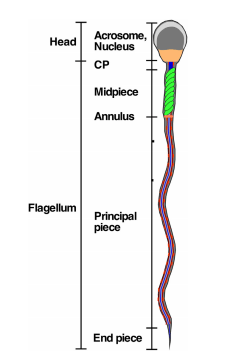
Illustration 3. Morphology of Spermatozoa (Gerton and Vadnalis, 2018).
REFERENCES
Auger, J. 2018. Semen Analysis. In: Skinner, M. K. (ed). Encyclopedia of Reproduction. Publisher Elsevier Science Publishing Co Inc, USA.
Garner D. L., and E. S. E. Hafez. 2000. Spermatozoa and Seminal Plasma. In: Hafez E. S. E. (ed). Reproduction in Farm Animals 7th Ed. Lippincott Williams & Wilkins, USA.
Gerton L. G., and M. L. Vadnalis. 2018. Structure of The Spermatozoon. In: Skinner, M. K. (ed). Encyclopedia of Reproduction. Publisher Elsevier Science Publishing Co Inc, USA.
Ismaya. 2014. Artificial Insemination Biotechnology in Cattle and Buffaloes. Gadjah Mada University Press, Yogyakarta.
Kretser D. M., P. Stanton, and L. O’Donnell. 2018. Structure/Cell Overview. In: Skinner, M. K. (ed). Encyclopedia of Reproduction. Publisher Elsevier Science Publishing Co Inc, USA.
Schenk J. L. 2018. Review: Principles of maximizing bull semen production at genetic centers. Animal. 12:142–147
Staub C. and L. Johnson. 2018. Review: Spermatogenesis in the bull. Animal. 12: 27–35
Susilawati, T.. 2011. Spermatology. UB Press, Malang.
Toelihere, M. R.. 1993. Artificial Insemination in Livestock. Angkasa Publishing, 3rd edition, Bandung.